Bursitis Top Of Foot Treatment Method
Overview
There are over 150 bursae in the human body. Usually bursae are present from birth, but they may form in response to repeated pressure. Each sac contains a small amount of synovial fluid, a clear liquid that acts as a lubricant. Inflammation causes pain on movement. The most common site for bursitis to occur is the shoulder (subdeltoid), but it also is seen in the elbows (olecranon), hips (trochanteric), knees, heels (Achilles), and toes. The affected area may be referred to as "frozen," because movement is so limited. In the knee there are four bursae, and all can become inflamed with overuse.
Causes
A rapid increase in physical activity levels or thinning of the heel?s protective fat pad are factors that may contribute to infracalcaneal bursitis. Other possible causes of infracalcaneal bursitis include blunt force trauma, acute or chronic infection, and arthritic conditions. The following factors may increase your risk of experiencing bursitis, including infracalcaneal bursitis. Participating in contact sports. Having a previous history of bursitis in any joint. Poor conditioning. Exposure to cold weather. Heel striking when running, especially in conventional running shoes with heel elevation.
Symptoms
What are the symptoms of heel bursitis? pain, swelling, tenderness, redness, and/or warmth at either the bottom of the heel or top of the heel, depending on the degree of swelling, pain may be a dull ache or substantial enough to cause limping, running, jumping, and walking activities may exacerbate pain, wearing poorly fitting, tight, or high-heeled shoes may exacerbate pain.
Diagnosis
Obtaining a detailed history from the patient is important in diagnosing calcaneal bursitis. The following complaints (which the physician should ask about during the subjective examination) are commonly reported by patients.
Other inquiries that the physician should make include the following. The clinician should ask about the patient's customary footwear (whether, for example, it includes high-heeled shoes or tight-fitting athletic shoes). The patient should be asked specifically about any recent change in footwear, such as whether he/she is wearing new athletic shoes or whether the patient has made a transition from flat shoes to high heels or vice versa. Individuals who have been accustomed to wearing high-heeled shoes on a long-term basis may find that switching to flat shoes causes increased stretch and irritation of the Achilles tendon and the associated bursae. The specifics of a patient's activity level should be ascertained, including how far the patient runs and, in particular, whether the individual is running with greater intensity than before or has increased the distance being run. The history of any known or suspected underlying rheumatologic conditions, such as gout, rheumatoid arthritis, or seronegative spondyloarthropathies, should be obtained.
Non Surgical Treatment
In addition to being aware of foot-wear and inserts, be sure to modify your activity level to reduce the pain initially. Taking non-steroidal anti-inflammatory drugs (NSAIDs) and icing twice a day for 20 minute periods can help reduce the swell that leads to heel pain. Cortisone injections (more powerful anti-inflammatory medications) can be considered if your symptoms are persistent. After the swelling and pain has receded, ask your podiatrist about working with a physical therapist to strengthen the affected area in order to avoid bursitis by using your muscles in a more safe and efficient manner. If all these treatment methods fail, surgery may be the best option to excise a painful bursa (note that this is in rare cases).
Surgical Treatment
Surgery. Though rare, particularly challenging cases of retrocalcaneal bursitis might warrant a bursectomy, in which the troublesome bursa is removed from the back of the ankle. Surgery can be effective, but operating on this boney area can cause complications, such as trouble with skin healing at the incision site. In addition to removing the bursa, a doctor may use the surgery to treat another condition associated with the retrocalcaneal bursitis. For example, a surgeon may remove a sliver of bone from the back of the heel to alter foot mechanics and reduce future friction. Any bone spurs located where the Achilles attaches to the heel may also be removed. Regardless of the conservative treatment that is provided, it is important to wait until all pain and swelling around the back of the heel is gone before resuming activities. This may take several weeks. Once symptoms are gone, a patient may make a gradual return to his or her activity level before their bursitis symptoms began. Returning to activities that cause friction or stress on the bursa before it is healed will likely cause bursitis symptoms to flare up again.
Treatment At Home For Hammer Toes
 Overview
A Hammer toes is a contracture-or bending-of the toe at the first joint of the digit, called the proximal interphalangeal joint. This bending causes the toe to appear like an upside-down hammertoes V when looked at from the side. Any toe can be involved, but the condition usually affects the second through fifth toes, known as the lesser digits. Hammertoes are more common to females than males.
Causes
Essentially, hammertoes are caused by an abnormal interworking of the bones, muscles, ligaments and tendons that comprise your feet. When muscles fail to work in a balanced manner, the toe joints can bend to form the hammertoe shape. If they remain in this position for an extended period, the muscles and tendons supporting them tighten and remain in that position. A common factor in development of hammertoe is wearing shoes that squeeze the toes or high heels that jam the toes into the front of the shoe. Most likely due to these factors, hammertoe occurs much more frequently in women than in men.
 Symptoms
Hammertoe and mallet toe feature an abnormal bend in the joints of one or more of your toes. Moving the affected toe may be difficult or painful. Corns and calluses can result from the toe rubbing against the inside of your shoes. See your doctor if you have persistent foot pain that affects your ability to walk properly.
Diagnosis
The treatment options vary with the type and severity of each hammer toe, although identifying the deformity early in its development is important to avoid surgery. Your podiatric physician will examine and X-ray the affected area and recommend a treatment plan specific to your condition.
Non Surgical Treatment
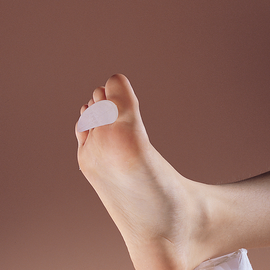
Apply a commercial, nonmedicated hammertoe pad around the bony prominence of the hammertoe. This will decrease pressure on the area. Wear a shoe with a deep toe box. If the hammertoe becomes inflamed and painful, apply ice packs several times a day to reduce swelling. Avoid heels more than two inches tall. A loose-fitting pair of shoes can also help protect the foot while reducing pressure on the affected toe, making walking a little easier until a visit to your podiatrist can be arranged. It is important to remember that, while this treatment will make the hammertoe feel better, it does not cure the condition. A trip to the podiatric physician?s office will be necessary to repair the toe to allow for normal foot function. Avoid wearing shoes that are too tight or narrow. Children should have their shoes properly fitted on a regular basis, as their feet can often outgrow their shoes rapidly. See your podiatric physician if pain persists.
Surgical Treatment
Probably the most frequent procedure performed is one called a Post or an Arthroplasty. In this case a small piece of bone is removed from the joint to straighten the toe. The toe is shortened somewhat, but there is still motion within the toe post-operatively. In other cases, an Arthrodesis is performed. This involves fusing the abnormally-contracted joint. The Taylor procedure fuses only the first joint in the toe, whereas the Lambrinudi procedure fuses both joints within the toe. Toes which have had these procedures are usually perfectly straight, but they take longer to heal and don't bend afterwards. A Hibbs procedure is a transfer of the toe's long extensor tendon to the top of the metatarsal bone. The idea of this procedure is to remove the deforming cause of the hammertoes (in this case, extensor substitution), but to preserve the tendon's function in dorsifexing the foot by reattaching it to the metatarsals. Fortunately, the Gotch (or Gotch and Kreuz) procedure--the removal of the base of the toe where it attaches to the foot, is done less frequently than in years past. The problem with this procedure is that it doesn't address the problem at the level of the deformity, and it causes the toe to become destabilized, often resulting in a toe that has contracted up and back onto the top of the foot. You can even have an Implant Arthroplasty procedure, where a small, false joint is inserted into place. There are several other procedures, as well.
 Prevention
To prevent a hammertoe, never squeeze your toes into shoes that force them to bend unnaturally. Those tendons can tighten up, and leave a permanent, claw-like bend in your toe. Always slip your feet into soft, roomy shoes that easily accommodate all of your toes. Stretching your toes can also help keep the tendons in the toes relaxed, and prevent a hammertoe. Use your hands to gently straighten and stretch your toes or try to pick up objects with your toes, grabbing something from the floor, for example. Sitting on a blanket and using your toes to grab the ends with also relax your feet.
Hammer Toe Repair Pinning
 Overview
A Hammer toe is a deformity of the second, third or fourth toes. In this condition, the toe is bent at the middle joint, so that it resembles a hammer. Initially, hammer toes are flexible and can be corrected with simple measures but, if left untreated, they can become fixed and require surgery. People with hammer toe may have corns or calluses on the top of the middle joint of the toe or on the tip of the toe. They may also feel pain in their toes or feet and have difficulty finding comfortable shoes.
Causes
A hammer toe develops because of an abnormal balance of the muscles in the toes. This abnormal balance causes increased pressures on the tendons and joints of the toe, leading to its contracture. Heredity and trauma can also lead to the formation of a hammer toe. Arthritis is another factor, because the balance around the toe in people with arthritis is disrupted. Wearing shoes that are too tight and cause Hammer toe the toes to squeeze can also cause a hammer toe to form.
 Symptoms
The symptoms of a hammer toe are usually first noticed when a corn develops on the top of the toe and becomes painful, usually when wearing tight shoes. There may be a bursa under the corn or instead of a corn, depending on the pressure. Most of the symptoms are due to pressure from footwear on the toe. There may be a callus under the metatarsal head at the base of the toe. Initially a hammer toe is usually flexible, but when longstanding it becomes more rigid.
Diagnosis
Most health care professionals can diagnose hammertoe simply by examining your toes and feet. X-rays of the feet are not needed to diagnose hammertoe, but they may be useful to look for signs of some types of arthritis (such as rheumatoid arthritis) or other disorders that can cause hammertoe. If the deformed toe is very painful, your doctor may recommend that you have a fluid sample withdrawn from the joint with a needle so the fluid can be checked for signs of infection or gout (arthritis from crystal deposits).
Non Surgical Treatment
Apply a commercial, nonmedicated hammertoe pad around the bony prominence of the hammertoe. This will decrease pressure on the area. Wear a shoe with a deep toe box. If the hammertoe becomes inflamed and painful, apply ice packs several times a day to reduce swelling. Avoid heels more than two inches tall. A loose-fitting pair of shoes can also help protect the foot while reducing pressure on the affected toe, making walking a little easier until a visit to your podiatrist can be arranged. It is important to remember that, while this treatment will make the hammertoe feel better, it does not cure the condition. A trip to the podiatric physician?s office will be necessary to repair the toe to allow for normal foot function. Avoid wearing shoes that are too tight or narrow. Children should have their shoes properly fitted on a regular basis, as their feet can often outgrow their shoes rapidly. See your podiatric physician if pain persists.
Surgical Treatment
Hammer toe can be corrected by surgery if conservative measures fail. Usually, surgery is done on an outpatient basis with a local anesthetic. The actual procedure will depend on the type and extent of the deformity. After the surgery, there may be some stiffness, swelling and redness and the toe may be slightly longer or shorter than before. You will be able to walk, but should not plan any long hikes while the toe heals, and should keep your foot elevated as much as possible.
 Prevention
Some different things you can do to help prevent hammer toes from forming and progressing do exist. You can wear supportive shoes to help prevent deformities. Hammer toes are often times related to poor foot mechanics, particularly foot flattening. You can wear custom orthotics prescribed by a podiatrist; orthotics might prevent or slow the progression of hammer toes. You can also avoid shoes with narrow or pointed toe boxes that can compress your toes.
Hallux Abducto Valgus Bunion Deformity
Overview
 A bunion (hallux valgus) is a deformity of the base joint of the big toe. The cause is not clear in many cases. The deformity may cause the foot to rub on shoes, which may cause inflammation and pain. Good footwear is often all that is needed to ease symptoms. An operation to correct the deformity is an option if good footwear does not ease symptoms.
Causes
Shoes. The primary cause of bunions is the long term use of shoes, particularly tight-fitting shoes with pointed toes, or high heeled shoes. A study that examined people in cultures that do not wear shoes found no cases of bunions. Genetic. People who have misaligned toes or feet, are flatfooted with feet that roll inwards (over pronation), excessive flexibility of ligaments, abnormal bone structure, or have mechanical instability in the big toe joint are more susceptible to bunions. This is especially common when bunions occur in children or young adults. Injuries or other trauma (sprains, fractures or nerve injuries), neuromuscular disorders (polio or Charcot-Marie-Tooth disease), or limb-length discrepancies (one leg longer than the other). Repetitive stresses to the foot. Bunions are common in ballet dancers and in a few sports. Arthritis.
Symptoms
With Bunions, a person will have inflammation, swelling, and soreness on the side surface of the big toe. Corns most commonly are tender cone-shaped patches of dry skin on the top or side of the toes. Calluses will appear on high-pressure points of the foot as thick hardened patches of skin.
Diagnosis
Looking at the problem area on the foot is the best way to discover a bunion. If it has the shape characteristic of a bunion, this is the first hint of a problem. The doctor may also look at the shape of your leg, ankle, and foot while you are standing, and check the range of motion of your toe and joints by asking you to move your toes in different directions A closer examination with weight-bearing X-rays helps your doctor examine the actual bone structure at the joint and see how severe the problem is. A doctor may ask about the types of shoes you wear, sports or activities (e.g., ballet) you participate in, and whether or not you have had a recent injury. This information will help determine your treatment.
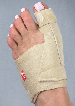
Non Surgical Treatment
The first step in the treatment of Bunions, Corns, and Calluses is determining what is causing the problem. Check with your family doctor or Podiatrist (foot doctor) to find the best solution for your ailment. You can reduce the risk of Bunions, Corns, and Calluses by following these simple instructions. Wear properly-fitting footwear, socks, and stockings (not too tight or too loose). Wear footwear with a wide toe box (toe area). Wear footwear or arch supports which provide proper support, weight distribution, and shock absorption. Maintain a healthy weight. For early-stage Bunions, soaking your feet in warm water can provide temporary relief. 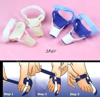
Surgical Treatment
The main goal of surgery is to realign the big toe joint in order to relieve symptoms, correct deformity and restore function. Surgery to remove a bunion is known as a bunionectomy. There are many variations of this operation and the type of surgery performed will vary depending on factors such as the degree of deformity, the strength of the bones, the person's age and the surgeon?s preferred approach. Most surgery involves the removal of the bony outgrowth (exostosis) and the realignment of the bones of the joint. Soft tissue structures such as the ligaments and tendons may be repositioned and the bursa may be removed. The insertion of screws and pins may be required to stabilise the bones in their new, realigned position.
Prevention
Bunions often become painful if they are allowed to progress. But not all bunions progress. Many bunion problems can be managed without surgery. In general, bunions that are not painful do not need surgical correction. For this reason, orthopaedic surgeons do not recommend "preventive" surgery for bunions that do not hurt; with proper preventive care, they may never become a problem.
Overpronation Of The Foot
Overview
Overpronation is the exaggerated inward rolling of the foot and ankle, which can lead to a collapsed arch and flat feet. Many people overpronate and do not even realize it; one way to tell is to simply look at the foot and see how it is placed on the ground when standing in a neutral position. Another way is to wet the bottom of the foot and step on a piece of paper. If the entire imprint of the foot is shown, it means you overpronate.
Causes
There may be several possible causes of over pronation. The condition may begin as early as birth. However, there are several more common explanations for the condition. First, wear and tear on the muscles throughout the foot, either from aging or repetitive strain, causes the muscles to weaken, thereby causing the foot to turn excessively inward. Also, standing or walking on high heels for an extended period of time also places strain and pressure on the foot which can weaken the tissue. Lastly, shoes play a very common factor in the development of over pronation. Shoes that fail to provide adequate support through the arch commonly lead to over pronation.
Symptoms
People with overpronation may suffer from pain in the knees, hips, and low back. Overpronation itself does not necessarily cause any other symptoms but is a contributing factor of many foot conditions such as Plantar Facsiitis, Heel Spur Syndrome, Posterior Tibialis Tendon Rupture or Tendonitis, Hallux Valgus, Bunion Deformities, Metatarsalgia, Hallux Limitus or Hallux Rigidus, Hammer Toes, and Morton?s Neuroma.
Diagnosis
Look at the wear on your shoes and especially running trainers; if you overpronate it's likely the inside of your shoe will be worn down (or seem crushed if they're soft shoes) from the extra strain.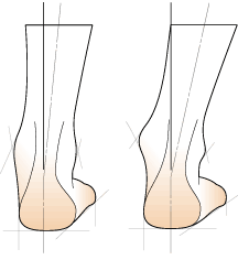
Non Surgical Treatment
If pronation is diagnosed before the age of five it can usually be treated in such a manner that the bones and joints will be aligned properly as growth continues. This may prevent the arch from collapsing, as well as allowing the muscles of the leg to enter the foot without twisting. With proper and early treatment, the foot will not turn out at the ankle, and the child?s gait will improve. Treatment for pronation in children may include: night braces, custom-made orthotics, and exercises. These treatments usually continue until growth is complete, and then the adult may need to wear custom-made orthotics to prevent the pronation from returning (the foot, as every other part of our body, tends to return to its original form if preventive measures are not taken). One side note: frequently, pediatricians will wait too long, hoping that the child will ?outgrow? the problem. By the time they realize that the child?s feet will not improve, it is too late to change the foot. In these cases, custom-made orthotics is used to prevent the pronation from becoming worse.
Prevention
Duck stance: Stand with your heels together and feet turned out. Tighten the buttock muscles, slightly tilt your pelvis forwards and try to rotate your legs outwards. You should feel your arches rising while you do this exercise.
Calf stretch:Stand facing a wall and place hands on it for support. Lean forwards until stretch is felt in the calves. Hold for 30 seconds. Bend at knees and hold for a further 30 seconds. Repeat 5 times.
Golf ball:While drawing your toes upwards towards your shins, roll a golf ball under the foot between 30 and 60 seconds. If you find a painful point, keep rolling the ball on that spot for 10 seconds.
Big toe push: Stand with your ankles in a neutral position (without rolling the foot inwards). Push down with your big toe but do not let the ankle roll inwards or the arch collapse. Hold for 5 seconds. Repeat 10 times. Build up to longer times and fewer repetitions.
Ankle strengthener: Place a ball between your foot and a wall. Sitting down and keeping your toes pointed upwards, press the outside of the foot against the ball, as though pushing it into the wall. Hold for 5 seconds and repeat 10 times.
Arch strengthener: Stand on one foot on the floor. The movements needed to remain balanced will strengthen the arch. When you are able to balance for 30 seconds, start doing this exercise using a wobble board.
Does Severs Disease Necessitate Surgery Treatment?
Overview
Sever?s Disease (calcaneal aphophysitis) is not really a disease, but more a repetitious strain injury. This is the most common cause of kids heel pain seen at Podiatry Care. Active children in football, soccer, basketball, netball and tennis with this type of foot pain complain of pain in the region of the heel bone particularly after exercise. In severe cases, children will complain of pain during exercise as well. It is a frequent cause of heel pain in children, particularly in the very active child. It is most often seen in children between the ages of 8 to 15 years as the growth plate is not fully developed or calcified at this time.
Causes
Overuse and stress on the heel bone through participation in sports is a major cause of calcaneal apophysitis. The heel?s growth plate is sensitive to repeated running and pounding on hard surfaces, resulting in muscle strain and inflamed tissue. For this reason, children and adolescents involved in soccer, track, or basketball are especially vulnerable. Other potential causes of calcaneal apophysitis include obesity, a tight Achilles tendon, and biomechanical problems such as flatfoot or a high-arched foot.
Symptoms
Patients with Severs disease typically experience pain that develops gradually in the back of the heel or Achilles region. In less severe cases, patients may only experience an ache or stiffness in the heel that increases with rest (especially at night or first thing in the morning). This typically occurs following activities which require strong or repetitive contraction of the calf muscles, such as running (especially uphill) or during sports involving running, jumping or hopping. The pain associated with this condition may also warm up with activity in the initial stages of the condition. As the condition progresses, patients may experience symptoms that increase during activity and affect performance. Pain may also increase when performing a calf stretch or heel raise (i.e. rising up onto tip toes). In severe cases, patients may walk with a limp, have difficulty putting their heel down, or be unable to weight bear on the affected leg. Pain may also increase on firmly touching the affected region and occasionally a bony lump may be palpable or visible at the back of the heel. This condition typically presents gradually overtime and can affect either one or both lower limbs.
Diagnosis
Sever?s disease is diagnosed based on a doctor?s physical examination of the lower leg, ankle, and foot. If the diagnosis is in question, the doctor may order x-rays or an MRI to determine if there are other injuries that may be causing the heel pain.
Non Surgical Treatment
See a doctor, who can diagnose the injury and recommend appropriate treatment options. It will be beneficial to rest the affected heel, and to regularly ice the affected area for the first few days. Anti-inflammatory pain medication can reduce pain and swelling, but first check with your doctor. As the pain diminishes a physical therapist can assist with a program of rehabilitation, incorporating stretching and strengthening exercises focused on the calf, shin and hamstring muscles. For a period after the injury has healed the doctor may advise on changes to your training routines that seek to lessen the strain on the heels. Orthotics are often recommended for your shoes in order to correct any biomechanical problems or lend extra support to the heels.
Pain In Left Foot Outside Arch
Overview
Plantar fasciitis refers to an inflammation of the plantar fascia, a thick, fibrous band running along the sole of the foot. Such inflammation results from direct injury to the plantar fascia, usually, repeated trauma to the tissue where the fascia attaches to the calcaneus or heel bone. The plantar fascia is critical in maintaining the foot?s complex arch system, also playing a role in balance and fine control of certain phases of the athlete?s gait. Injury to the plantar fascia is particularly painful and disabling for runners and can often prove stubbornly resistant to treatment. Rehabilitation is frequently a lengthy and frustrating process. For these reasons, care should be taken where possible to avoid such injury by means of preventative exercises and sensitivity to early warning signs.

Causes
The most common cause of arch pain is plantar fasciitis. Plantar fasciitis is the name that describes inflammation of the fibrous band of tissue that connects the heel to the toes. Symptoms of plantar fasciitis include pain early in the morning and pain with long walks or prolonged standing. Arch pain early in the morning is due to the plantar fascia becoming contracted and tight as you sleep through the night. When awakening and walking in the morning, the fascia is still tight and prone to irritation when stretched. When walking or standing for long periods, the plantar fascia becomes inflamed and painful. Treatment of plantar fasciitis is best accomplished with some simple stretching exercises, anti-inflammatory medications, and inserts for your shoes.
Symptoms
Go to a podiatrist at the first sign of symptoms. Besides pain on the bottom of the foot, additional symptoms may include burning sensation in arch, difficulty standing on tiptoes, inflammation, more pain after sleeping or resting, redness, heat, localized pain in the ball of the foot, sharp or shooting pain in the toes, pain that increases when toes are flexed, tingling or numbness in the toes, aching, pain that increases when walking barefoot, pain that increases when walking on hard surfaces, pain the increases when standing (putting weight on your feet) or moving around and decreases when immobile, skin Lesions, it?s important to get a proper diagnosis and treatment plan. Let?s go over the possible causes of the pain.
Diagnosis
Magnetic Resonance Imaging (MRI) can show tendon injury and inflammation but cannot be relied on with 100% accuracy and confidence. The technique and skill of the radiologist in properly positioning the foot with the MRI beam are critical in demonstrating the sometimes obscure findings of tendon injury around the ankle. Magnetic Resonance Imaging (MRI) is expensive and is not necessary in most cases to diagnose posterior tibial tendon injury. Ultrasound has also been used in some cases to diagnose tendon injury, but this test again is usually not required to make the initial diagnosis.
Non Surgical Treatment
High arches that are flexible do not require any treatment. In cases where there is pain, shoe modifications such as an arch insert or support insole can help to relieve pain during walking. Custom orthotic devices can be given that fit into the shoe and provide stability and cushioning effect. Your doctor may recommend a brace to help keep the foot and ankle stable. In severe cases, surgery is performed to flatten the foot. Any coexisting nerve disorders are also treated.

Surgical Treatment
Although most patients with plantar fasciitis respond to non- surgical treatment, a small percentage of patients may require surgery. If, after several months of non-surgical treatment, you continue to have heel pain, surgery will be considered. Your foot and ankle surgeon will discuss the surgical options with you and determine which approach would be most beneficial. No matter what kind of treatment you undergo for plantar fasciitis, the underlying causes that led to this condition may remain. Therefore, you will need to continue with preventive measures. Wearing supportive shoes, stretching, and using custom orthotic devices are the mainstays of long-term treatment for plantar fasciitis.
Prevention
Strap the arches into the anatomically correct positions with athletic tape and leave them like this for some time. If the fallen arches are an issue with the muscular structure, this may give the muscles an opportunity to strengthen. This is definitely not a fallen arches cure all the time but it can help prevent it more times than not. Ask a doctor or physical therapists to show you how to do this taping. Find shoes that fit. This may require that you get your foot measured and molded to ensure that the shoe will fit. Shoes that are too big, too tight or too short, may not directly cause the fallen arches, but they can assist with the damage to the area. These shoes should have thick cushioning inside and have plenty of room for your toes. Walk without shoes as much as possible. Shoes directly assist with weakening and distorting the arches of the feet so going without shoes can actually help strengthen your arches and prevent fallen arches. Walking on hard and bumpy surfaces barefooted makes the muscles in your feet strengthen in order to prevent injury. It is a coping mechanism by your body. Insert heel cups or insoles into the shoes that you wear the most. Many people wear uncomfortable shoes to work and these are the same shoes that cause their arches the most problems. Inserting the heel cups and insoles into these shoes can prevent fallen arches from occurring. Many people place these inserts into all their shoes to ensure support. Ask a medical professional, either your doctor or a physical therapist, about daily foot exercises that may keep the arches stronger than normal. Many times, you can find exercises and stretches on the Internet on various websites. Curling your toes tightly and rotating your feet will help strengthen your longitudinal arches. Relax your feet and shake them for a minute or so before you do any arch exercises. This will loosen the muscles in your feet that stay tight due to normal daily activities. Wear rigid soled sandals whenever possible to provide a strong support for your arches. Wooden soled sandals are the best ones if available. Walk or jog on concrete as much as you can. This will create a sturdy support for your arches. Running or walking in sandy areas or even on a treadmill, does not give rigid support. Instead, these surfaces absorb the step, offering no support for arches.
Stretching Exercises
Massage therapy is a great way to loosen muscles and help improve mobility in in your feet. As many people with foot pain have discovered, tight muscles in your legs or back can lead to tense foot muscles. All those muscles are connected, so tension in your back can cause tension in your legs which can pull the tendons in your feet and cause stiffness and pain. Getting acupuncture or a professional full body massage are probably the best ways to deal with this, but there are also some simple tricks you can do at home to help keep muscles limber. These are great for loosening up and improving circulation, both before and after exercise. Place a tennis ball under the arch of your bare foot and roll it around, stretching the muscles in your foot and promoting blood flow. You can also roll the ball under your calves and upper legs to work out stiffness and knots. If you feel the tennis ball is too easy, try a lacrosse ball for deeper massaging. This is also demonstrated in the exercise video above. Use a foam roller, those big overpriced rolls of foam that are now available in every department and sporting goods store are fantastic for self-massage (why a roll of foam costs $30 is beyond us, but they do work wonders-our advice is to not waste money on the more expensive fancy grooved ones because even the simplest rollers work great). The exercises you can do with foam rollers seem to be endless, and there are literally hundreds of free videos online showing how to use them to massage every part of your body. Here's one we picked out that specifically targets foot and leg muscles related to arches and plantar fasciitis.
Plantar fasciitis refers to an inflammation of the plantar fascia, a thick, fibrous band running along the sole of the foot. Such inflammation results from direct injury to the plantar fascia, usually, repeated trauma to the tissue where the fascia attaches to the calcaneus or heel bone. The plantar fascia is critical in maintaining the foot?s complex arch system, also playing a role in balance and fine control of certain phases of the athlete?s gait. Injury to the plantar fascia is particularly painful and disabling for runners and can often prove stubbornly resistant to treatment. Rehabilitation is frequently a lengthy and frustrating process. For these reasons, care should be taken where possible to avoid such injury by means of preventative exercises and sensitivity to early warning signs.
Causes
The most common cause of arch pain is plantar fasciitis. Plantar fasciitis is the name that describes inflammation of the fibrous band of tissue that connects the heel to the toes. Symptoms of plantar fasciitis include pain early in the morning and pain with long walks or prolonged standing. Arch pain early in the morning is due to the plantar fascia becoming contracted and tight as you sleep through the night. When awakening and walking in the morning, the fascia is still tight and prone to irritation when stretched. When walking or standing for long periods, the plantar fascia becomes inflamed and painful. Treatment of plantar fasciitis is best accomplished with some simple stretching exercises, anti-inflammatory medications, and inserts for your shoes.
Symptoms
Go to a podiatrist at the first sign of symptoms. Besides pain on the bottom of the foot, additional symptoms may include burning sensation in arch, difficulty standing on tiptoes, inflammation, more pain after sleeping or resting, redness, heat, localized pain in the ball of the foot, sharp or shooting pain in the toes, pain that increases when toes are flexed, tingling or numbness in the toes, aching, pain that increases when walking barefoot, pain that increases when walking on hard surfaces, pain the increases when standing (putting weight on your feet) or moving around and decreases when immobile, skin Lesions, it?s important to get a proper diagnosis and treatment plan. Let?s go over the possible causes of the pain.
Diagnosis
Magnetic Resonance Imaging (MRI) can show tendon injury and inflammation but cannot be relied on with 100% accuracy and confidence. The technique and skill of the radiologist in properly positioning the foot with the MRI beam are critical in demonstrating the sometimes obscure findings of tendon injury around the ankle. Magnetic Resonance Imaging (MRI) is expensive and is not necessary in most cases to diagnose posterior tibial tendon injury. Ultrasound has also been used in some cases to diagnose tendon injury, but this test again is usually not required to make the initial diagnosis.
Non Surgical Treatment
High arches that are flexible do not require any treatment. In cases where there is pain, shoe modifications such as an arch insert or support insole can help to relieve pain during walking. Custom orthotic devices can be given that fit into the shoe and provide stability and cushioning effect. Your doctor may recommend a brace to help keep the foot and ankle stable. In severe cases, surgery is performed to flatten the foot. Any coexisting nerve disorders are also treated.
Surgical Treatment
Although most patients with plantar fasciitis respond to non- surgical treatment, a small percentage of patients may require surgery. If, after several months of non-surgical treatment, you continue to have heel pain, surgery will be considered. Your foot and ankle surgeon will discuss the surgical options with you and determine which approach would be most beneficial. No matter what kind of treatment you undergo for plantar fasciitis, the underlying causes that led to this condition may remain. Therefore, you will need to continue with preventive measures. Wearing supportive shoes, stretching, and using custom orthotic devices are the mainstays of long-term treatment for plantar fasciitis.
Prevention
Strap the arches into the anatomically correct positions with athletic tape and leave them like this for some time. If the fallen arches are an issue with the muscular structure, this may give the muscles an opportunity to strengthen. This is definitely not a fallen arches cure all the time but it can help prevent it more times than not. Ask a doctor or physical therapists to show you how to do this taping. Find shoes that fit. This may require that you get your foot measured and molded to ensure that the shoe will fit. Shoes that are too big, too tight or too short, may not directly cause the fallen arches, but they can assist with the damage to the area. These shoes should have thick cushioning inside and have plenty of room for your toes. Walk without shoes as much as possible. Shoes directly assist with weakening and distorting the arches of the feet so going without shoes can actually help strengthen your arches and prevent fallen arches. Walking on hard and bumpy surfaces barefooted makes the muscles in your feet strengthen in order to prevent injury. It is a coping mechanism by your body. Insert heel cups or insoles into the shoes that you wear the most. Many people wear uncomfortable shoes to work and these are the same shoes that cause their arches the most problems. Inserting the heel cups and insoles into these shoes can prevent fallen arches from occurring. Many people place these inserts into all their shoes to ensure support. Ask a medical professional, either your doctor or a physical therapist, about daily foot exercises that may keep the arches stronger than normal. Many times, you can find exercises and stretches on the Internet on various websites. Curling your toes tightly and rotating your feet will help strengthen your longitudinal arches. Relax your feet and shake them for a minute or so before you do any arch exercises. This will loosen the muscles in your feet that stay tight due to normal daily activities. Wear rigid soled sandals whenever possible to provide a strong support for your arches. Wooden soled sandals are the best ones if available. Walk or jog on concrete as much as you can. This will create a sturdy support for your arches. Running or walking in sandy areas or even on a treadmill, does not give rigid support. Instead, these surfaces absorb the step, offering no support for arches.
Stretching Exercises
Massage therapy is a great way to loosen muscles and help improve mobility in in your feet. As many people with foot pain have discovered, tight muscles in your legs or back can lead to tense foot muscles. All those muscles are connected, so tension in your back can cause tension in your legs which can pull the tendons in your feet and cause stiffness and pain. Getting acupuncture or a professional full body massage are probably the best ways to deal with this, but there are also some simple tricks you can do at home to help keep muscles limber. These are great for loosening up and improving circulation, both before and after exercise. Place a tennis ball under the arch of your bare foot and roll it around, stretching the muscles in your foot and promoting blood flow. You can also roll the ball under your calves and upper legs to work out stiffness and knots. If you feel the tennis ball is too easy, try a lacrosse ball for deeper massaging. This is also demonstrated in the exercise video above. Use a foam roller, those big overpriced rolls of foam that are now available in every department and sporting goods store are fantastic for self-massage (why a roll of foam costs $30 is beyond us, but they do work wonders-our advice is to not waste money on the more expensive fancy grooved ones because even the simplest rollers work great). The exercises you can do with foam rollers seem to be endless, and there are literally hundreds of free videos online showing how to use them to massage every part of your body. Here's one we picked out that specifically targets foot and leg muscles related to arches and plantar fasciitis.